|
 Electrolyte Monitoring during Furosemide Therapy: a Practical Guide
Understanding Electrolyte Shifts with Loop Diuretics
A sudden bout of edema can make the decision to start furosemide feel urgent; clinicians must balance rapid symptom relief against invisible electrolyte upheaval. Awareness of how loop diuretics alter renal handling of sodium, potassium, magnesium and calcium turns a risky blindfold into a manageable checklist. Mechanistically, loops inhibit the Na+-K+-2Cl− cotransporter in the thick ascending limb, causing natriuresis and volume contraction. Loss of lumen-positive potential reduces paracellular reabsorption of magnesium and calcium, while increased distal sodium delivery drives potassium wasting and predisposes to metabolic alkalosis. Electrolyte shifts can be immediate (within hours) or accumulate over days. Hyponatremia often reflects free water retention or excessive natriuresis, whereas hypokalemia and hypomagnesemia commonly cause muscle cramps, arrhythmias, and refractory potassium loss unless magnesium is corrected. | Electrolyte | Change | | Na+ | ↓/variable | | K+ | ↓ | | Mg2+ | ↓ |
Baseline Tests and Risk Stratification before Starting Therapy
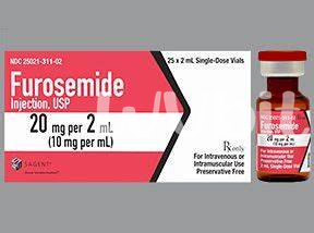
Imagine the first clinic visit as a checkpoint: review history, baseline chemistry, renal function, and ECG when indicated, because furosemide can unmask vulnerabilities. Document volume status, medications that affect electrolytes, and factors like age or heart failure that raise risk. A focused baseline panel guides safe initiation. Stratify patients into low- and high-risk groups and tailor monitoring: repeat labs within days for high risk, within weeks for stable patients. Consider magnesium and potassium supplements preemptively in vulnerable individuals, and coordinate with specialists when chronic kidney disease or recurrent hospitalizations complicate care.
Monitoring Schedule: Frequency and Practical Timing Tips
Start monitoring early: baseline electrolytes, renal function and weight inform frequency, then adjust with clinical response. For most patients beginning furosemide, check serum sodium, potassium and creatinine within 3–7 days, then again at two weeks; in high‑risk cases or rapid diuresis, measure within 24–48 hours. Time labs before the morning dose to avoid transient post‑dose shifts. Longitudinal monitoring should become individualized: stable outpatients may move to monthly checks for three months, then quarterly, while those on concomitant renin‑angiotensin blockers or with heart failure need closer surveillance. Encourage patients to report dizziness, cramps or reduced urine output, and coordinate lab timing with clinic visits to improve adherence and decision making and medication reconciliation at each visit.
Recognizing and Interpreting Common Lab Abnormalities Clinically
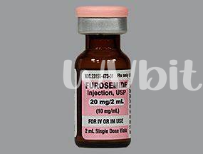
A patient with leg swelling looks relieved when lab results arrive; clinicians should read serum sodium, potassium, chloride, bicarbonate and creatinine with that relief tempered by awareness of diuretic effects. Loop diuretics like furosemide cause concentrated shifts — natriuresis, kaliuresis and volume contraction — which change values rapidly. Interpretation hinges on trends and clinical context: rising creatinine with falling blood pressure suggests overdiuresis, while isolated low potassium without renal impairment points to urinary losses. Assess osmolar status, urine electrolytes when needed, and correlate with symptoms such as muscle cramps, weakness or confusion. Acting on labs requires graded responses: modest electrolyte changes may call for oral supplements and diet adjustment, but severe hyponatremia or hypokalemia mandates prompt replacement and reassessment of diuretic dose. Clear documentation and repeat testing after interventions close the loop between numbers and patient outcomes for safer ongoing care planning.
Management Strategies for Hyponatremia, Hypokalemia, Hypomagnesemia
A clear plan transforms electrolyte chaos into manageable steps. When using furosemide, anticipate sodium and potassium shifts, explain risks, and set thresholds for intervention so both clinician and patient know when to act promptly confidently. Start with baseline labs and frequent checks after dose changes. For hyponatremia, restrict free water and consider cautious hypertonic saline for severe symptoms. For hypokalemia, prefer oral supplements and potassium-sparing agents when feasible with monitoring. Hypomagnesemia often coexists with low potassium; correct magnesium to enable potassium repletion. Use oral magnesium oxide for mild deficits, intravenous magnesium sulfate for severe or symptomatic cases, and reassess levels within 24 to 48 hours. Educate patients about symptom recognition, diet, and avoiding interacting drugs like NSAIDs or laxatives. Schedule timely lab checks after dose adjustments. Escalate care for arrhythmia, persistent electrolyte loss, or failure to improve despite replacement therapy.
| Issue | Quick Action |
|---|
| Hyponatremia | Fluid restriction / Na+ prn |
| Hypokalemia | Oral K+ or IV if severe |
| Hypomagnesemia | Oral Mg2+ or IV MgSO4 |
Patient Education, Diet, Drug Interactions, and Follow-up
Begin with clear, patient-centered explanations of expected effects and red flags: muscle cramps, lightheadedness, palpitations and reduced urine output. Encourage a simple symptom diary. Stress the importance of reporting fainting or severe weakness promptly. Diet advice should focus on consistent potassium intake from fruits and vegetables, avoiding excessive sodium restriction unless otherwise directed; discuss magnesium-rich foods like nuts and leafy greens. Tailor guidance for older adults and those with comorbidities. Review interacting medications such as NSAIDs, ACE inhibitors, digoxin and aminoglycosides. Counsel about alcohol and over-the-counter supplements, and advise bringing all medicines to appointments. Arrange baseline and repeat labs, set clear follow-up timing, and explain when urgent review is needed. Trusted resources: MedlinePlus and PubChem
|